Esophageal Squamous Cell Carcinoma Stage IB
| Title: | Esophageal Squamous Cell Carcinoma Stage IB |
|---|---|
| Description: |
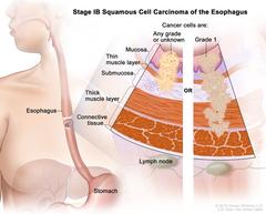
Stage IB squamous cell carcinoma of the esophagus; drawing shows the esophagus and stomach. A two-panel inset shows the layers of the esophagus wall: the mucosa layer, thin muscle layer, submucosa layer, thick muscle layer, and connective tissue layer. The lymph nodes are also shown. The left panel shows cancer cells that are any grade or of an unknown grade in the mucosa layer, thin muscle layer, and submucosa layer. The right panel shows grade 1 cancer cells in the mucosa layer, thin muscle layer, submucosa layer, and thick muscle layer. Stage IB squamous cell carcinoma of the esophagus. Cancer has spread into the mucosa layer, thin muscle layer, or submucosa layer of the esophagus wall. The cancer cells are any grade or the grade is not known; OR cancer has spread into the thick muscle layer of the esophagus wall. The cancer cells are grade 1. Grade 1 cancer cells look more like normal cells under a microscope and grow and spread more slowly than grade 2 and 3 cancer cells. |
| Topics/Categories: |
Anatomy -- Digestive/Gastrointestinal System Cancer Types -- Esophageal Cancer Cells or Tissue -- Abnormal Cells or Tissue Staging |
| Type: | Color, Medical Illustration (JPEG format) |
| Source: | National Cancer Institute |
| Creator: | Terese Winslow (Illustrator) |
| AV Number: | CDR752731 |
| Date Created: | July 18, 2019 |
| Date Added: | October 28, 2014 |
| Reuse Restrictions: |
Yes - This image is copyright protected. Any use of this image is subject to prevailing copyright laws. U.S. Government has reuse rights. Please contact the rights holder of this image for permission requests.
Rights holder: Terese Winslow Email: terese@teresewinslow.com |