Ciliary Pocket Membrane
| Title: | Ciliary Pocket Membrane |
|---|---|
| Description: |
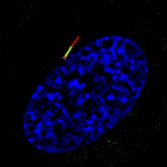
Understanding the mechanisms regulating transport of signaling molecules to and from the cilia is important for developing drugs targeted against ciliary-associated cancer pathways. The ciliary membrane is continuous with the plasma membrane through a specialized pocket-like membrane referred to as the ciliary pocket. Here, structured illumination microscopy (SIM) imaging of RPE cells shows the ciliary membrane and ciliary pocket. The membrane reshaping protein EHD1 (green) specifically accumulates at the ciliary pocket membrane while the Hedgehog pathways receptor Smoothened localizes to the ciliary membrane (red). The base of the cilia is marked by the mother centriole distal appendage protein CEP164 (pink) and nuclei (blue). This image was originally submitted as part of the 2015 NCI Cancer Close Up project. |
| Topics/Categories: | Cells or Tissue -- Normal Cells or Tissue |
| Type: | Color, Photo (JPEG format) |
| Source: | NCI Center for Cancer Research |
| Creator: | Quanlong Lu, Christopher Westlake |
| Date Created: | 2014 |
| Date Added: | April 9, 2015 |
| Reuse Restrictions: | None - This image is in the public domain and can be freely reused. Please credit the source and, where possible, the creator listed above. |