Esophageal Adenocarcinoma Stage IIIB (1)
| Title: | Esophageal Adenocarcinoma Stage IIIB (1) |
|---|---|
| Description: |
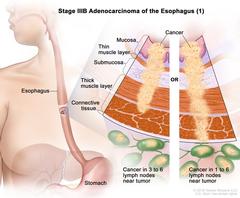
Stage IIIB adenocarcinoma of the esophagus (1); drawing shows the esophagus and stomach. An inset shows the layers of the esophagus wall: the mucosa layer, thin muscle layer, submucosa layer, thick muscle layer, and connective tissue layer. The left panel shows cancer in the mucosa layer, thin muscle layer, submucosa layer, and thick muscle layer and in 3 lymph nodes near the tumor. The right panel shows cancer in the mucosa layer, thin muscle layer, submucosa layer, thick muscle layer, and connective tissue layer and in 4 lymph nodes near the tumor. Stage IIIB adenocarcinoma of the esophagus (1). Cancer has spread into the thick muscle layer of the esophagus wall. Cancer is found in 3 to 6 lymph nodes near the tumor; OR cancer has spread into the connective tissue layer of the esophagus wall. Cancer is found in 1 to 6 lymph nodes near the tumor. |
| Topics/Categories: |
Anatomy -- Digestive/Gastrointestinal System Anatomy -- Lymphatic System Cancer Types -- Esophageal Cancer Cells or Tissue -- Abnormal Cells or Tissue Staging |
| Type: | Color, Medical Illustration (JPEG format) |
| Source: | National Cancer Institute |
| Creator: | Terese Winslow (Illustrator) |
| AV Number: | CDR762234 |
| Date Created: | August 13, 2019 |
| Date Added: | March 4, 2015 |
| Reuse Restrictions: |
Yes - This image is copyright protected. Any use of this image is subject to prevailing copyright laws. U.S. Government has reuse rights. Please contact the rights holder of this image for permission requests.
Rights holder: Terese Winslow Email: terese@teresewinslow.com |