Cilia and Ciliary Pocket
| Title: | Cilia and Ciliary Pocket |
|---|---|
| Description: |
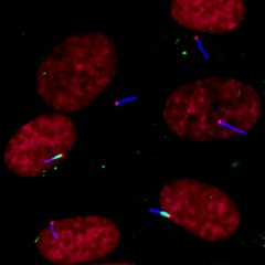
Signal transduction pathways allow molecules inside of a cell to be altered by molecules on the outside. Some pathways work via cilia on the cell surface. Defects in ciliary formation and function are linked to several human diseases, including cancer. Epifluorescence imaging shows a cluster of human retinal pigment epithelium (RPE) cells displaying primary cilia. Cells were serum starved for 24 hours before imaging to induce cilia formation. Cilia (blue, anti-acetylated tubulin), EHD1 (green, ciliary pocket), centrosome (red, anti-Cep164) and nuclei (dark red). This image was originally submitted as part of the 2015 NCI Cancer Close Up project. |
| Topics/Categories: | Cells or Tissue -- Normal Cells or Tissue |
| Type: | Color, Photo (JPEG format) |
| Source: | NCI Center for Cancer Research |
| Creator: | Quanlong Lu, Christopher Westlake |
| Date Created: | 2013 |
| Date Added: | April 8, 2015 |
| Reuse Restrictions: | None - This image is in the public domain and can be freely reused. Please credit the source and, where possible, the creator listed above. |