Fibroblast
| Title: | Fibroblast |
|---|---|
| Description: |
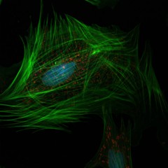
A confocal microscopy image of a fibroblast showing the nucleus (blue), mitochondria (red), and actin cytoskeleton (green). As is evident from their large number of mitochondria, fibroblasts are very metabolically active, continuously synthesizing elements of the extracellular matrix and collagen. Tissue damage is a major trigger for the activation of fibroblasts from fibrocytes, and so fibroblasts play an important role in wound healing. Fibroblast assays are currently being studied as a means of predicting how normal tissue may respond to radiation therapy in cancer patients and others. This image was originally submitted as part of the 2015 NCI Cancer Close Up project. |
| Topics/Categories: |
Cells or Tissue -- Normal Cells or Tissue Treatment -- Radiation Therapy |
| Type: | Color, Photo (JPEG format) |
| Source: | NCI Center for Cancer Research |
| Creator: | Guy Jones, Matthew Dreher, Brad Wood |
| Date Created: | 2015 |
| Date Added: | April 9, 2015 |
| Reuse Restrictions: | None - This image is in the public domain and can be freely reused. Please credit the source and, where possible, the creator listed above. |