Brain, Medial, Child, Anatomy-HP
| Title: | Brain, Medial, Child, Anatomy-HP |
|---|---|
| Description: |
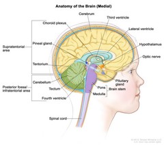
Drawing of the inside of the brain showing the supratentorial area (the upper part of the brain) and the posterior fossa/infratentorial area (the lower back part of the brain). The supratentorial area contains the cerebrum, lateral ventricle, third ventricle, choroid plexus, hypothalamus, pineal gland, pituitary gland, and optic nerve. The posterior fossa/infratentorial area contains the cerebellum, tectum, fourth ventricle, and brain stem (pons and medulla). The tentorium and spinal cord are also shown. Anatomy of the inside of the brain. The supratentorial area (the upper part of the brain) contains the cerebrum, lateral ventricle and third ventricle (with cerebrospinal fluid shown in blue), choroid plexus, hypothalamus, pineal gland, pituitary gland, and optic nerve. The posterior fossa/infratentorial area (the lower back part of the brain) contains the cerebellum, tectum, fourth ventricle, and brain stem (pons and medulla). The tentorium separates the supratentorium from the infratentorium. |
| Topics/Categories: |
Anatomy -- Nervous System People -- Child |
| Type: | Color, Medical Illustration (JPEG format) |
| Source: | National Cancer Institute |
| Creator: | Terese Winslow (Illustrator) |
| AV Number: | CDR748659 |
| Date Created: | November 10, 2015 |
| Date Added: | November 25, 2013 |
| Reuse Restrictions: |
Yes - This image is copyright protected. Any use of this image is subject to prevailing copyright laws. U.S. Government has reuse rights. Please contact the rights holder of this image for permission requests.
Rights holder: Terese Winslow Email: terese@teresewinslow.com |