Bile Duct, Extrahepatic, Anatomy
| Title: | Bile Duct, Extrahepatic, Anatomy |
|---|---|
| Description: |
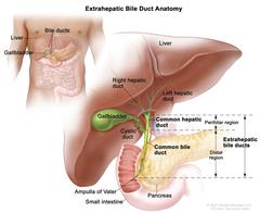
Anatomy of the extrahepatic bile ducts; drawing shows the extrahepatic bile ducts, including the common hepatic duct (perihilar region) and the common bile duct (distal region). Also shown are the liver, right and left hepatic ducts, gallbladder, cystic duct, pancreas, ampulla of Vater, and small intestine. Anatomy of the extrahepatic bile ducts. Extrahepatic bile ducts are small tubes that carry bile from the liver and gallbladder to the small intestine. They are made up of the common hepatic duct (perihilar region) and the common bile duct (distal region). Bile is made in the liver and flows through the common hepatic duct and the cystic duct to the gallbladder, where it is stored. When food is being digested, bile is released from the gallbladder and flows through the common bile duct, pancreas, and ampulla of Vater into the small intestine. Anatomy of the extrahepatic bile ducts. |
| Topics/Categories: | Anatomy -- Digestive/Gastrointestinal System |
| Type: | Color, Medical Illustration (JPEG format) |
| Source: | National Cancer Institute |
| Creator: | Terese Winslow (Illustrator) |
| AV Number: | CDR659742 |
| Date Created: | July 31, 2023 |
| Date Added: | April 15, 2011 |
| Reuse Restrictions: |
Yes - This image is copyright protected. Any use of this image is subject to prevailing copyright laws. U.S. Government has reuse rights. Please contact the rights holder of this image for permission requests.
Rights holder: Terese Winslow Email: terese@teresewinslow.com |