Skin With Melanocyte Anatomy-HP
| Title: | Skin With Melanocyte Anatomy-HP |
|---|---|
| Description: |
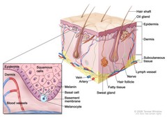
Schematic representation of normal skin; drawing shows normal skin anatomy, including the epidermis, dermis, hair follicles, sweat glands, hair shafts, veins, arteries, fatty tissue, nerves, lymph vessels, oil glands, and subcutaneous tissue. The pullout shows a close-up of the squamous cell and basal cell layers of the epidermis, the basement membrane in between the epidermis and dermis, and the dermis with blood vessels. Melanin is shown in the cells. A melanocyte is shown in the layer of basal cells at the deepest part of the epidermis. Schematic representation of normal skin. The relatively avascular epidermis houses basal cell keratinocytes and squamous epithelial keratinocytes, the source cells for BCC and SCC, respectively. Melanocytes are also present in normal skin and serve as the source cell for melanoma. The separation between epidermis and dermis occurs at the basement membrane zone, located just inferior to the basal cell keratinocytes. |
| Topics/Categories: |
Anatomy -- Skin Cells or Tissue -- Normal Cells or Tissue |
| Type: | Color, Medical Illustration (JPEG format) |
| Source: | National Cancer Institute |
| Creator: | Terese Winslow (Illustrator) |
| AV Number: | CDR624959 |
| Date Created: | September 24, 2008 |
| Date Added: | March 8, 2010 |
| Reuse Restrictions: |
Yes - This image is copyright protected. Any use of this image is subject to prevailing copyright laws. U.S. Government has reuse rights. Please contact the rights holder of this image for permission requests.
Rights holder: Terese Winslow Email: terese@teresewinslow.com |