Ultrasound, Transvaginal
| Title: | Ultrasound, Transvaginal |
|---|---|
| Description: |
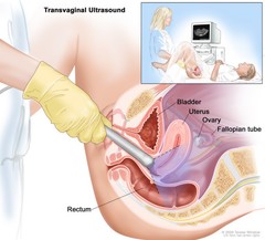
Transvaginal ultrasound; drawing shows a side view of the female reproductive anatomy during a transvaginal ultrasound procedure. An ultrasound probe (a device that makes sound waves that bounce off tissues inside the body) is shown inserted into the vagina. The bladder, uterus, right fallopian tube, and right ovary are also shown. The inset shows the diagnostic sonographer (a person trained to perform ultrasound procedures) examining a woman on a table, and a computer screen shows an image of the patient's internal tissues. Transvaginal ultrasound. An ultrasound probe connected to a computer is inserted into the vagina and is gently moved to show different organs. The probe bounces sound waves off internal organs and tissues to make echoes that form a sonogram (computer picture). |
| Topics/Categories: |
Anatomy -- Gynecologic Test or Procedure -- Imaging Procedures |
| Type: | Color, Medical Illustration (JPEG format) |
| Source: | National Cancer Institute |
| Creator: | Terese Winslow (Illustrator) |
| AV Number: | CDR618018 |
| Date Created: | November 9, 2012 |
| Date Added: | March 8, 2010 |
| Reuse Restrictions: |
Yes - This image is copyright protected. Any use of this image is subject to prevailing copyright laws. U.S. Government has reuse rights. Please contact the rights holder of this image for permission requests.
Rights holder: Terese Winslow Email: terese@teresewinslow.com |