Respiratory System Anatomy
| Title: | Respiratory System Anatomy |
|---|---|
| Description: |
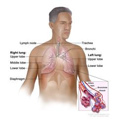
Respiratory system anatomy; drawing shows the right lung with the upper, middle, and lower lobes, the left lung with the upper and lower lobes, and the trachea, bronchi, lymph nodes, and diaphragm. An inset shows the bronchioles, alveoli, artery, and vein. Anatomy of the respiratory system showing the trachea, the right and left lungs and their lobes, and the bronchi. The lymph nodes and the diaphragm are also shown. Oxygen is inhaled into the lungs and passes through the alveoli (the tiny air sacs at the end of the bronchioles) and into the bloodstream (see inset), where it travels to the tissues throughout the body. Anatomy of the respiratory system. |
| Topics/Categories: | Anatomy -- Respiratory/Thoracic System |
| Type: | Color, Medical Illustration (JPEG format) |
| Source: | National Cancer Institute |
| Creator: | Terese Winslow (Illustrator) |
| AV Number: | CDR466533 |
| Date Created: | March 27, 2024 |
| Date Added: | September 2, 2008 |
| Reuse Restrictions: |
Yes - This image is copyright protected. Any use of this image is subject to prevailing copyright laws. U.S. Government has reuse rights. Please contact the rights holder of this image for permission requests.
Rights holder: Terese Winslow Email: terese@teresewinslow.com |