Ductal Carcinoma Histology
| Title: | Ductal Carcinoma Histology |
|---|---|
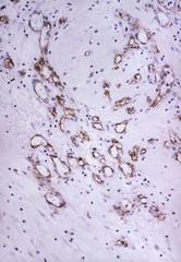
| Description: | (a) cross-section of infiltrating ductal carcinoma of the breast with blood vessels at the periphery of tumor. Magnification x100. (b) cross-section of infiltrating ductal carcinoma of the breast with vessels at the periphery of tumor. Magnification x200. (c) cross section of infiltrating ductal carcinoma of the breast with a small foci of breast cancer cells in which cd34 antibody has stained blood vessels and basement membrane. Magnification x100. (d) cross-section of infiltrating ductal carcinoma of breast shows intense blood vessel proliferation in stromal tissue adjacent to the malignant tissue. Magnification x200. |
| Topics/Categories: |
Cancer Types -- Breast Cancer Cells or Tissue -- Abnormal Cells or Tissue |
| Type: | Color, Photo (JPEG format) |
| Source: | National Cancer Institute |
| Creator: | Unknown Photographer |
| AV Number: | AV-9500-4293 |
| Date Created: | 1995 |
| Date Added: | January 1, 2001 |
| Reuse Restrictions: | None - This image is in the public domain and can be freely reused. Please credit the source and, where possible, the creator listed above. |