Cancer Cells: Death (Step 2)
| Title: | Cancer Cells: Death (Step 2) |
|---|---|
| Description: |
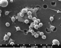
Step-two in a six-step sequence of the death of a cancer cell. A cancer cell has migrated through the holes of a matrix coated membrane from the top to the bottom, simulating natural migration of a invading cancer cell between, and sometimes through, the vascular endothelium. A buffy coat containing red blood cells, lymphocytes and macrophages is added to the bottom of the membrane. A group of macrophages identify the cancer cell as foreign matter and start to stick to the cancer cell, which still has its spikes (step 2). Photo magnification: x4,000. See also: http://visuals.nci.nih.gov/details.cfm?imageid=2369 http://visuals.nci.nih.gov/details.cfm?imageid=2370 http://visuals.nci.nih.gov/details.cfm?imageid=2371 http://visuals.nci.nih.gov/details.cfm?imageid=2372 http://visuals.nci.nih.gov/details.cfm?imageid=2373 |
| Topics/Categories: | Cells or Tissue -- Abnormal Cells or Tissue |
| Type: | B&W, Photo (JPEG format) |
| Source: | Dr. Raowf Guirguis. National Cancer Institute |
| Creator: | Susan Arnold (Photographer) |
| AV Number: | AV-8810-3685-B |
| Date Created: | October 1988 |
| Date Added: | January 1, 2001 |
| Reuse Restrictions: | None - This image is in the public domain and can be freely reused. Please credit the source and, where possible, the creator listed above. |