| Description: |
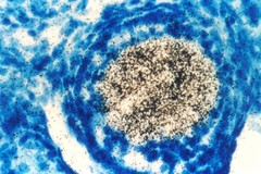
A technique called in situ hybridization shows whether a gene is actively expressed in cells, and also provides clues to the gene's function. This technique has helped identify activated oncogenes in cancer cells, and their normal counterparts in normal cells, in many different species. In this photograph, a labeled DNA segment (a known oncogene) has been put into a mouse oocyte, a cell that develops into a mature egg cell. The labeled DNA has paired with (or hybridized to) multiple copies of RNA in the mouse oocyte. The presence of this RNA (shown here as black dots inside the nucleus of the immature cell) shows that the normal cellular counterpart of the oncogene is active, suggesting that it is critical for normal germ cell development. Expression of genes is manifested by the production of RNA transcripts within cells. Hybridization histochemistry (in situ hybridization) permits localization of these transcripts with cellular or greater resolution. Furthermore, the relative amounts of transcripts detected within different tissues or the same tissues under different states (e.g., physiological or developmental) may be quantified.
|