| Description: |
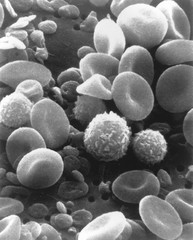
This is a scanning electron microscope image from normal circulating human blood. One can see red blood cells, several white blood cells including lymphocytes, a monocyte, a neutrophil, and many small disc-shaped platelets. Red cells are nonnucleated, and contain hemoglobin, containing iron an important protein which allows the cell to carry oxygen to other parts of the body. They also carry away carbon dioxide from the lungs. The infection-fighting white blood cells, are classified in 2 main groups: granular and agranular. Granulocytes are formed in bone marrow, agranulocytes are produced by lymph nodes and spleen. There are two types of agranulocytes: lymphocytes, fight disease by producing antibodies and thus destroying foreign material, and monocytes. Platelets are tiny cells formed in bone marrow and are necessary for blood clotting.
|