| Description: |
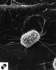
This is a scanning electron micrograph of a dividing cell, cultured from chinese hampster ovary tissue (cho). The light micrograph (inset) of the same mitotic cell reveals that it's in the anaphase stage when the darkly stained chromosomes move to opposite poles of the cell prior to cell cleavage. The surface of this cell, seen in the scanning electron micrograph image, is covered with small fingerlike projections called microvilli; this surface appearance is typical, but not definitive, for cultured cells in anaphase.
|