| Title: |
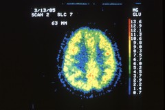
PET Scan of a Normal Brain |
| Description: |
Position emission tomography (PET) of cerebral glucose utilization in a normal individual. This tomogram is through the upper part of the cerebral hemisphere. Note discrimination between gray matter (yellow-red color) and white matter (green-blue color) which uses less glucose.
|
| Topics/Categories: |
Test or Procedure -- Imaging Procedures |
| Type: |
Color, Other (JPEG format) |
| Source: |
Dr. Giovanni Dichiro, Neuroimaging Section, National Institute of Neurological Disorders and Stroke |
| Creator: |
Unknown Photographer |
| AV Number: |
AV-0000-3635 |
| Date Created: |
Unknown |
| Date Added: |
January 1, 2001 |
| Reuse Restrictions: |
None - This image is in the public domain and can be freely reused. Please credit the source and, where possible, the creator listed above.
|