Lymphedema-HP
| Title: | Lymphedema-HP |
|---|---|
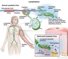
| Description: | Lymphedema; the top part of the drawing shows normal lymphatic flow. An arrow is used to show intravascular fluid flowing through an artery, an arteriole, and a capillary bed, where the fluid leaks out into the interstitial space around the cells and then exits through the venules. Also shown is interstitial fluid, large proteins, and cells entering a lymphatic vessel to maintain normal interstitial pressure. The fluid in the lymphatic vessel is called lymph. Also shown is the inside structure of a lymph node attached to the lymphatic vessel with arrows showing how the lymph moves into and out of the lymph node. The lymphatic ducts in the neck area of a female figure are also shown. The figure’s left arm is red and swollen. There is a pull-out from the swollen arm showing a top layer of red, hardened skin and an inset box showing obstructed lymphatic flow. A damaged lymphatic vessel resulting in increased interstitial pressure and a build-up of large proteins, cellular debris, macrophages, and lymphocytes are shown. Large fat cells and fibrosis are also shown in the inset box. |
| Topics/Categories: |
Anatomy -- Lymphatic System Anatomy -- Skin |
| Type: | Color, Medical Illustration (JPEG format) |
| Source: | National Cancer Institute |
| Creator: | Terese Winslow (Illustrator) |
| AV Number: | CDR812467 |
| Date Created: | August 21, 2023 |
| Date Added: | December 20, 2023 |
| Reuse Restrictions: |
Yes - This image is copyright protected. Any use of this image is subject to prevailing copyright laws. U.S. Government has reuse rights. Please contact the rights holder of this image for permission requests.
Rights holder: Terese Winslow Email: terese@teresewinslow.com |