Cervix Anatomy
| Title: | Cervix Anatomy |
|---|---|
| Description: |
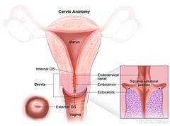
Drawing of the anatomy of the cervix showing the internal OS, endocervical canal, endocervix, ectocervix, and external OS. The uterus and vagina are also shown. There is also a pullout that shows a close up of the squamocolumnar junction (the area where the endocervix and ectocervix meet) and the cells that line the endocervix and ectocervix. Anatomy of the cervix. The cervix is the lower, narrow end of the uterus that connects the uterus to the vagina. It is made up of the internal OS (the opening between the cervix and the upper part of the uterus), the endocervix (the inner part of the cervix that forms the endocervical canal), the ectocervix (the outer part of the cervix that opens into the vagina) and the external OS (the opening between the cervix and vagina). The area where the endocervix and ectocervix meet is called the squamocolumnar junction, which contains both glandular cells (column-shaped cells that make mucus) from the endocervix and squamous cells (thin, flat cells) from the ectocervix. The squamocolumnar junction is sometimes referred to as the transformation zone. |
| Topics/Categories: | Anatomy -- Gynecologic |
| Type: | Color, Medical Illustration (JPEG format) |
| Source: | National Cancer Institute |
| Creator: | Terese Winslow (Illustrator) |
| AV Number: | CDR810405 |
| Date Created: | August 9, 2023 |
| Date Added: | July 21, 2023 |
| Reuse Restrictions: |
Yes - This image is copyright protected. Any use of this image is subject to prevailing copyright laws. U.S. Government has reuse rights. Please contact the rights holder of this image for permission requests.
Rights holder: Terese Winslow Email: terese@teresewinslow.com |