Esophagus Anatomy, Child
| Title: | Esophagus Anatomy, Child |
|---|---|
| Description: |
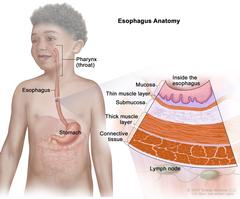
Anatomy of the esophagus; drawing shows the pharynx (throat), esophagus, and stomach. A pullout shows the mucosa layer, thin muscle layer, submucosa layer, thick muscle layer, and connective tissue layer of the esophagus wall. The lymph nodes are also shown. Anatomy of the esophagus. The esophagus is a hollow, muscular tube that moves food and liquid from the pharynx (throat) to the stomach. The wall of the esophagus is made up of several layers of tissue, including the mucosa layer, thin muscle layer, submucosa layer, thick muscle layer, and connective tissue layer. |
| Topics/Categories: |
Anatomy -- Digestive/Gastrointestinal System Anatomy -- Lymphatic System |
| Type: | Color, Medical Illustration (JPEG format) |
| Source: | National Cancer Institute |
| Creator: | Terese Winslow (Illustrator) |
| AV Number: | CDR804898 |
| Date Created: | May 17, 2021 |
| Date Added: | June 15, 2021 |
| Reuse Restrictions: |
Yes - This image is copyright protected. Any use of this image is subject to prevailing copyright laws. U.S. Government has reuse rights. Please contact the rights holder of this image for permission requests.
Rights holder: Terese Winslow Email: terese@teresewinslow.com |