Eye Anatomy
| Title: | Eye Anatomy |
|---|---|
| Description: |
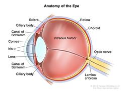
Anatomy of the eye; drawing shows the sclera, ciliary body, canal of Schlemm, cornea, iris, lens, vitreous humor, retina, choroid, optic nerve, and lamina cribrosa. Anatomy of the eye showing the sclera, ciliary body, canal of Schlemm, cornea, iris, lens, vitreous humor, retina, choroid, optic nerve, and lamina cribrosa. The vitreous humor is a gel that fills the center of the eye. |
| Topics/Categories: | Anatomy -- Eye |
| Type: | Color, Medical Illustration (JPEG format) |
| Source: | National Cancer Institute |
| Creator: | Terese Winslow (Illustrator) |
| AV Number: | CDR798975 |
| Date Created: | August 9, 2019 |
| Date Added: | August 21, 2019 |
| Reuse Restrictions: |
Yes - This image is copyright protected. Any use of this image is subject to prevailing copyright laws. U.S. Government has reuse rights. Please contact the rights holder of this image for permission requests.
Rights holder: Terese Winslow Email: terese@teresewinslow.com |