VHL Renal Tumors, Bilateral
| Title: | VHL Renal Tumors, Bilateral |
|---|---|
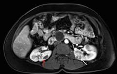
| Description: |
Axial view of an individual’s midsection showing tumors in both kidneys. The left kidney has a tumor with a dark cystic component and the right kidney has a predominantly solid tumor. Vista de un corte transversal del cuerpo donde se observan tumores en ambos riñones. En el riñón izquierdo se ve un tumor con un componente quístico oscuro y en el riñón derecho se ve un tumor predominantemente sólido. VHL-associated RCCs are characteristically multifocal and bilateral and present as a combined cystic and solid mass. Red arrow indicates a lesion with a solid and cystic component and white arrow indicates a predominantly solid lesion. Los carcinomas de células renales asociados con la enfermedad de VHL suelen ser multifocales y bilaterales. Se presentan como una masa con componentes quísticos y sólidos. La flecha roja señala una lesión con un componente sólido y un componente quístico, la flecha blanca señala una lesión predominantemente sólida. |
| Topics/Categories: |
Anatomy -- Genitourinary Test or Procedure -- Imaging Procedures |
| Type: | B&W, Photo (JPEG format) |
| Source: | National Cancer Institute |
| Creator: | Brian Shuch, MD, Yale University School of Medicine (Photographer) |
| AV Number: | CDR778858 |
| Date Created: | February 16, 2016 |
| Date Added: | February 22, 2016 |
| Reuse Restrictions: | None - This image is in the public domain and can be freely reused. Please credit the source and, where possible, the creator listed above. |