Ovarian Tumor Microenvironment
| Title: | Ovarian Tumor Microenvironment |
|---|---|
| Description: |
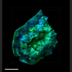
In this image from a mouse model of ovarian cancer, optically cleared tumor excised from a murine SKOV tumor seeded with CD63+ cells reveals a high-resolution landscape of the three-dimensional tumor-stromal interfaces that comprise the tumor microenvironment (TME). Second harmonic signal (blue) and autofluorescent /GFP signals (green) demonstrates the interplay of collagen II fibrils and vessels generated from angiogenesis. The use of optical tissue clearing has the potential to greatly improve researchers' ability to assess the anatomic, structural, and cellular constituents that govern metastatic colonization in the TME at a single-cell resolution. This image was originally submitted as part of the 2016 NCI Cancer Close Up project. |
| Topics/Categories: |
Cancer Types -- Ovarian Cancer Cells or Tissue -- Abnormal Cells or Tissue |
| Type: | Color, Photo (JPEG format) |
| Source: | National Cancer Institute \ Comprehensive Cancer Center of Wake Forest Univ. |
| Creator: | Chris Booth, Kyle Cowdrick, Frank C. Marini |
| Date Created: | July 2013 |
| Date Added: | April 11, 2016 |
| Reuse Restrictions: | None - This image is in the public domain and can be freely reused. Please credit the source and, where possible, the creator listed above. |