Breast Tumor Cells Using Microtentacles
| Title: | Breast Tumor Cells Using Microtentacles |
|---|---|
| Description: |
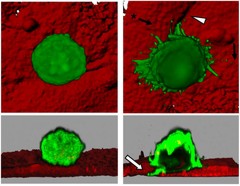
3D confocal microscopy datasets of live human mammary epithelial cells (green) using microtentacles (black arrows) to penetrate the junctions (white arrowhead) between blood vessel endothelial cells (red). Cross sections (lower panels) show that microtentacles extend through cell-cell junctions to penetrate underneath the endothelial monolayer (white arrow). A nontumorigenic mammary epithelial cell remains on the endothelial surface (left panels), while a cell that has undergone epithelial-to-mesenchymal transition (right panels) generates microtentacles that penetrate endothelial layers. This image was originally submitted as part of the 2016 NCI Cancer Close Up project. |
| Topics/Categories: |
Cancer Types -- Breast Cancer Cells or Tissue -- Abnormal Cells or Tissue |
| Type: | Color, Photo (JPEG format) |
| Source: | National Cancer Institute \ Univ. of Maryland Greenebaum Cancer Center |
| Creator: | Stuart S. Martin |
| Date Created: | March 2010 |
| Date Added: | April 14, 2016 |
| Reuse Restrictions: | None - This image is in the public domain and can be freely reused. Please credit the source and, where possible, the creator listed above. |