Breast Tumor Microenvironment
| Title: | Breast Tumor Microenvironment |
|---|---|
| Description: |
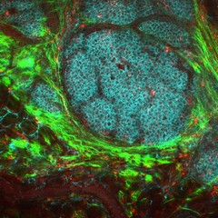
This image of a breast cancer tumor and its microenvironment was obtained from a live mouse model using multiphoton microscopy and endogenous fluorescence. That is, the image was obtained without any fluorophores, stains, or dyes, using only the metabolic co-factors of NADH and FAD, which are already inside of cells, along with second harmonic generation to see collagen. This technique has important clinical potential for patients who require label-free imaging, and may lead to more effective diagnoses and treatments. Tumor cells display in cyan, macrophages in red, collagen fibers in green. This image was originally submitted as part of the 2016 NCI Cancer Close Up project and selected for exhibit. |
| Topics/Categories: |
Cancer Types -- Breast Cancer Cells or Tissue -- Abnormal Cells or Tissue |
| Type: | Color, Photo (JPEG format) |
| Source: | National Cancer Institute \ Carbone Cancer Center at the Univ. of Wisconsin |
| Creator: | Joseph Szulczewski, David Inman, Kevin Eliceiri, and Patricia Keely |
| Date Created: | January 2016 |
| Date Added: | April 11, 2016 |
| Reuse Restrictions: | None - This image is in the public domain and can be freely reused. Please credit the source and, where possible, the creator listed above. |