Bile Duct, Intrahepatic, Anatomy
| Title: | Bile Duct, Intrahepatic, Anatomy |
|---|---|
| Description: |
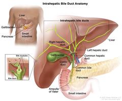
Anatomy of the intrahepatic bile ducts; drawing shows the liver and the intrahepatic bile ducts, which include the right and left hepatic ducts. Also shown is the common hepatic duct, gallbladder, cystic duct, common bile duct, pancreas, ampulla of Vater, and small intestine. An inset shows a cross section of a liver lobule with a network of bile ductules leading into a bile duct. Anatomy of the intrahepatic bile ducts. Intrahepatic bile ducts are a network of small tubes that carry bile inside the liver. The smallest ducts, called ductules, come together to form the right and left hepatic ducts, which lead out of the liver. The two ducts join outside the liver and form the common hepatic duct. The cystic duct from the gallbladder joins the common hepatic duct to form the common bile duct. The common bile duct passes through the pancreas and ends in the small intestine. Bile is made by the liver and stored in the gallbladder. When food is being digested, bile is released from the gallbladder and passes through the common bile duct, pancreas, and ampulla of Vater into the small intestine. Anatomy of the intrahepatic bile ducts. |
| Topics/Categories: | Anatomy -- Digestive/Gastrointestinal System |
| Type: | Color, Medical Illustration (JPEG format) |
| Source: | National Cancer Institute |
| Creator: | Terese Winslow (Illustrator) |
| AV Number: | CDR765897 |
| Date Created: | July 31, 2023 |
| Date Added: | December 4, 2015 |
| Reuse Restrictions: |
Yes - This image is copyright protected. Any use of this image is subject to prevailing copyright laws. U.S. Government has reuse rights. Please contact the rights holder of this image for permission requests.
Rights holder: Terese Winslow Email: terese@teresewinslow.com |