Vascular Invasion by Cancer Cells
| Title: | Vascular Invasion by Cancer Cells |
|---|---|
| Description: |
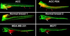
Zebrafish are a useful animal model in which to study the characteristics and behavior of human cancer cells. Here, a variety of human cell lines (labeled in red) were injected in zebrafish embryos: normal human cells (breast cells 1 and 2); human tumor cells (adenoid cystic carcinoma of salivary gland, or ACC); non-metastatic tumor cells (MCF7, ACC PDX, and MDA-MB231). The metastatic tumor cells, but not normal or non-metastatic tumor cells, exhibit vascular invasion in zebrafish within 5-7 days post injection. The animals' vascular system is labeled green; white arrows indicate site of human cell injection; red arrows indicate the invasion of tumor cells; and red arrows point to the cells that have extravasated. This image was originally submitted as part of the 2016 NCI Cancer Close Up project. |
| Topics/Categories: | Cells or Tissue -- Abnormal Cells or Tissue |
| Type: | Color, Photo (JPEG format) |
| Source: | National Cancer Institute \ Georgetown Lombardi Comprehensive Cancer Center |
| Creator: | Seema Agarwal |
| Date Created: | 2015 |
| Date Added: | April 11, 2016 |
| Reuse Restrictions: | None - This image is in the public domain and can be freely reused. Please credit the source and, where possible, the creator listed above. |