Detecting Abnormal Cells in the Cervix
| Title: | Detecting Abnormal Cells in the Cervix |
|---|---|
| Description: |
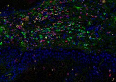
This image of cellular infiltrate in mucous membrane cells of the cervix visualizes a panel of biomarkers indicating abnormal expansion or a growth. The biomarkers detected include CD3 (yellow); CD8 (red); PD1 (pink) and PDL1 (green). The blue color represents the cell nucleus. The intensity of each fluorescent signal can be measured and interpreted as the amount of that particular protein present in the tissue sample. This image indicates the presence of different inflammatory cells and immune regulatory cells present in the cellular infiltrate. This image was originally submitted as part of the 2016 NCI Cancer Close Up project. |
| Topics/Categories: |
Cancer Types -- Cervical Cancer Cells or Tissue -- Abnormal Cells or Tissue |
| Type: | Color, Photo (JPEG format) |
| Source: | National Cancer Institute \ Tisch Cancer Institute at the Mount Sinai School of Medicine |
| Creator: | Tin Htwe Thin |
| Date Created: | April 2015 |
| Date Added: | April 11, 2016 |
| Reuse Restrictions: | None - This image is in the public domain and can be freely reused. Please credit the source and, where possible, the creator listed above. |