Intestinal Epithelial Cell-Cell Boundary
| Title: | Intestinal Epithelial Cell-Cell Boundary |
|---|---|
| Description: |
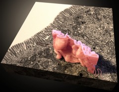
Three-dimensional structure of the intersection of two cells lining the small intestine (orange), shown with the stack of electron microscopy images that the structure was derived from. Data from focused ion beam scanning electron microscopy (FIB-SEM). See also www.electron.nci.nih.gov/. |
| Topics/Categories: | Cells or Tissue -- Normal Cells or Tissue |
| Type: | Color, Illustration (JPEG format) |
| Source: | National Cancer Institute (NCI) |
| Creator: | Amy Moran (NLM), Sriram Subramaniam |
| Date Created: | September 2014 |
| Date Added: | December 3, 2015 |
| Reuse Restrictions: | None - This image is in the public domain and can be freely reused. Please credit the source and, where possible, the creator listed above. |