Ciliogenesis Progression
| Title: | Ciliogenesis Progression |
|---|---|
| Description: |
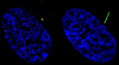
Defects in ciliary formation and function are linked to several human diseases, including cancer. Some cancer cells appear to have lost the ability to develop or maintain cilia, which may contribute to tumorigensis. Here, structured illumination microscopy imaging of RPE cells shows ciliogenesis progression. Ciliogenesis starts from docking pre-ciliary vesicles to the distal appendage of the mother centriole. Those docked vesicles fuse to form a larger ciliary vesicle and then extend to the ciliary membrane surrounding the growing microtubule-based axoneme. A GFP-labeled G protein-coupled receptor called Smoothened localizes to these docked vesicles and also the ciliary membrane. Left cell shows GFP-Smoothened (green) vesicles docked at the mother centriole/basal body distal appendage (marked by CEP164 in red). Right cell shows ciliary GFP-Smoothened localized on ciliary membrane. Nuclei (blue). This image was originally submitted as part of the 2015 NCI Cancer Close Up project. |
| Topics/Categories: | Cells or Tissue -- Normal Cells or Tissue |
| Type: | Color, Photo (JPEG format) |
| Source: | NCI Center for Cancer Research |
| Creator: | Quanlong Lu, Christopher Westlake |
| Date Created: | 2014 |
| Date Added: | April 9, 2015 |
| Reuse Restrictions: | None - This image is in the public domain and can be freely reused. Please credit the source and, where possible, the creator listed above. |