Carcinoma In Situ
| Title: | Carcinoma In Situ |
|---|---|
| Description: |
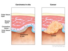
Two-panel drawing showing layers of tissue, including the outer tissue layer, muscle layers, connective tissue, and inner tissue layer. The left panel shows carcinoma in situ (abnormal cells) in the inner tissue layer. The right panel shows cancer cells spreading from the inner tissue layer to the connective tissue and muscle layers. Carcinoma in situ (CIS) is a group of abnormal cells that are found only in the place where they first formed in the body (see left panel). These abnormal cells may become cancer and spread to nearby normal tissue (see right panel). |
| Topics/Categories: | Cells or Tissue -- Abnormal Cells or Tissue |
| Type: | Color, Medical Illustration (JPEG format) |
| Source: | National Cancer Institute |
| Creator: | Terese Winslow (Illustrator) |
| AV Number: | CDR761914 |
| Date Created: | October 2, 2014 |
| Date Added: | October 28, 2014 |
| Reuse Restrictions: |
Yes - This image is copyright protected. Any use of this image is subject to prevailing copyright laws. U.S. Government has reuse rights. Please contact the rights holder of this image for permission requests.
Rights holder: Terese Winslow Email: terese@teresewinslow.com |