Brain, Medial, Child, Anatomy
| Title: | Brain, Medial, Child, Anatomy |
|---|---|
| Description: |
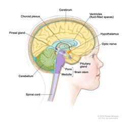
Drawing of the inside of the brain showing ventricles (fluid-filled spaces), choroid plexus, hypothalamus, pineal gland, pituitary gland, optic nerve, brain stem, cerebellum, cerebrum, medulla, pons, and spinal cord. Anatomy of the inside of the brain, showing the pineal and pituitary glands, optic nerve, ventricles (with cerebrospinal fluid shown in blue), and other parts of the brain. Anatomy of the inside of the brain, showing the pineal and pituitary glands, optic nerve, ventricles (with cerebrospinal fluid shown in blue), and other parts of the brain. The posterior fossa is the region below the tentorium, which separates the cortex from the cerebellum and essentially denotes the region containing the brain stem, cerebellum, and fourth ventricle. |
| Topics/Categories: |
Anatomy -- Nervous System People -- Child |
| Type: | Color, Medical Illustration (JPEG format) |
| Source: | National Cancer Institute |
| Creator: | Terese Winslow (Illustrator) |
| AV Number: | CDR689771 |
| Date Created: | November 17, 2010 |
| Date Added: | April 15, 2011 |
| Reuse Restrictions: |
Yes - This image is copyright protected. Any use of this image is subject to prevailing copyright laws. U.S. Government has reuse rights. Please contact the rights holder of this image for permission requests.
Rights holder: Terese Winslow Email: terese@teresewinslow.com |