Biopsy, Sentinel Lymph Node, Breast (3-Panel)
| Title: | Biopsy, Sentinel Lymph Node, Breast (3-Panel) |
|---|---|
| Description: |
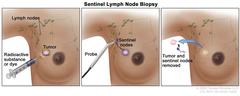
Sentinel lymph node biopsy of the breast; the first panel shows a radioactive substance and/or blue dye being injected near the tumor; the second panel shows that the injected material is followed visually and/or with a probe that detects radioactivity to find the sentinel nodes (the first lymph nodes to which cancer cells are likely to spread from a primary tumor); and the third panel shows the removal of the tumor and the sentinel nodes to check for cancer cells. Sentinel lymph node biopsy of the breast. A radioactive substance and/or blue dye is injected near the tumor (first panel). The injected material is followed visually and/or with a probe that detects radioactivity to find the sentinel nodes (the first lymph nodes to which cancer cells are likely to spread from a primary tumor) (second panel). The sentinel nodes are removed and checked for cancer cells (third panel). A sentinel lymph node biopsy is usually done at the same time the primary tumor is removed, but it can also be done before or after the tumor is removed. |
| Topics/Categories: |
Anatomy -- Breast Cancer Types -- Breast Cancer Test or Procedure -- Biopsy |
| Type: | Color, Medical Illustration (JPEG format) |
| Source: | National Cancer Institute |
| Creator: | Terese Winslow (Illustrator) |
| AV Number: | CDR661757 |
| Date Created: | October 24, 2022 |
| Date Added: | April 15, 2011 |
| Reuse Restrictions: |
Yes - This image is copyright protected. Any use of this image is subject to prevailing copyright laws. U.S. Government has reuse rights. Please contact the rights holder of this image for permission requests.
Rights holder: Terese Winslow Email: terese@teresewinslow.com |