Hypopharynx Anatomy
| Title: | Hypopharynx Anatomy |
|---|---|
| Description: |
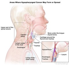
Drawing shows areas where hypopharyngeal cancer may form or spread, including the bone under the tongue (hyoid bone), cartilage around the thyroid and trachea, the thyroid, the trachea, and the esophagus. Also shown are the upper part of the spinal column, the carotid artery, lymph nodes in the neck, and lining of the chest cavity. An inset shows a cross section of the hypopharynx, larynx, esophagus, and trachea. Hypopharyngeal cancer forms in the tissues of the hypopharynx (the bottom part of the throat). It may spread to nearby tissues or to cartilage around the thyroid or trachea, the bone under the tongue (hyoid bone), the thyroid, the trachea, the larynx, or the esophagus. It may also spread to the lymph nodes in the neck, the carotid artery, the tissues around the upper part of the spinal column, the lining of the chest cavity, and to other parts of the body (not shown). |
| Topics/Categories: | Anatomy -- Respiratory/Thoracic System |
| Type: | Color, Medical Illustration (JPEG format) |
| Source: | National Cancer Institute |
| Creator: | Terese Winslow (Illustrator) |
| AV Number: | CDR780507 |
| Date Created: | May 10, 2017 |
| Date Added: | May 15, 2017 |
| Reuse Restrictions: |
Yes - This image is copyright protected. Any use of this image is subject to prevailing copyright laws. U.S. Government has reuse rights. Please contact the rights holder of this image for permission requests.
Rights holder: Terese Winslow Email: terese@teresewinslow.com |