Ovarian Cancer Stage I
| Title: | Ovarian Cancer Stage I |
|---|---|
| Description: |
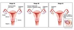
Three-panel drawing of stage IA, stage IB, and stage IC; each panel shows the ovaries, fallopian tubes, uterus, cervix, and vagina. The first panel (stage IA) shows cancer inside one ovary. The second panel (stage IB) shows cancer inside both ovaries. The third panel (stage IC) shows cancer inside both ovaries and (a) the tumor in the ovary on the left has ruptured (broken open), (b) there is cancer on the surface of the ovary on the right, and (c) there are cancer cells in the pelvic peritoneal fluid (inset). In stage IA, cancer is found inside a single ovary or fallopian tube. In stage IB, cancer is found inside both ovaries or fallopian tubes. In stage IC, cancer is found inside one or both ovaries or fallopian tubes and one of the following is true: (a) either the tumor or the capsule (outer covering) of the ovary has ruptured (broken open), or (b) cancer is also found on the surface of the ovary or fallopian tube, or (c) cancer cells are found in the pelvic peritoneal fluid. |
| Topics/Categories: |
Anatomy -- Gynecologic Cancer Types -- Ovarian Cancer Cells or Tissue -- Abnormal Cells or Tissue Staging |
| Type: | Color, Medical Illustration (JPEG format) |
| Source: | National Cancer Institute |
| Creator: | Terese Winslow (Illustrator) |
| AV Number: | CDR618025 |
| Date Created: | June 20, 2022 |
| Date Added: | March 8, 2010 |
| Reuse Restrictions: |
Yes - This image is copyright protected. Any use of this image is subject to prevailing copyright laws. U.S. Government has reuse rights. Please contact the rights holder of this image for permission requests.
Rights holder: Terese Winslow Email: terese@teresewinslow.com |