Biopsy, Fine Needle Aspiration (FNA), Lung
| Title: | Biopsy, Fine Needle Aspiration (FNA), Lung |
|---|---|
| Description: |
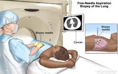
Fine-needle aspiration biopsy of the lung; drawing shows a patient lying on a table that slides through the computed tomography (CT) machine with an x-ray picture of a cross-section of the lung on a monitor above the patient. Drawing also shows a doctor using the x-ray picture to help place the biopsy needle through the chest wall and into the area of abnormal lung tissue. Inset shows a side view of the chest cavity and lungs with the biopsy needle inserted into the area of abnormal tissue. Fine-needle aspiration biopsy of the lung. The patient lies on a table that slides through the computed tomography (CT) machine, which takes x-ray pictures of the inside of the body. The x-ray pictures help the doctor see where the abnormal tissue is in the lung. A biopsy needle is inserted through the chest wall and into the area of abnormal lung tissue. A small piece of tissue is removed through the needle and checked under the microscope for signs of cancer. |
| Topics/Categories: |
Anatomy -- Respiratory/Thoracic System Cancer Types -- Lung Cancer Cells or Tissue -- Abnormal Cells or Tissue Test or Procedure -- Biopsy |
| Type: | Color, Medical Illustration (JPEG format) |
| Source: | National Cancer Institute |
| Creator: | Terese Winslow (Illustrator) |
| AV Number: | CDR531057 |
| Date Created: | September 27, 2022 |
| Date Added: | August 27, 2008 |
| Reuse Restrictions: |
Yes - This image is copyright protected. Any use of this image is subject to prevailing copyright laws. U.S. Government has reuse rights. Please contact the rights holder of this image for permission requests.
Rights holder: Terese Winslow Email: terese@teresewinslow.com |