Cancer Cells: Death (Step 3)
| Title: | Cancer Cells: Death (Step 3) |
|---|---|
| Description: |
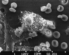
Step-three of a six-step sequence of the death of a cancer cell. A cancer cell has migrated through the holes of a matrix coated membrane from the top to the bottom, simulating natural migration of a invading cancer cell between, and sometimes through, the vascular endothelium. Macrophages begin to fuse with, and inject its toxins into, the cancer cell. The cell starts rounding up and loses its spikes (step 3). Photo magnification: x8,000. See also: http://visuals.nci.nih.gov/details.cfm?imageid=2369 http://visuals.nci.nih.gov/details.cfm?imageid=2370 http://visuals.nci.nih.gov/details.cfm?imageid=2371 http://visuals.nci.nih.gov/details.cfm?imageid=2372 http://visuals.nci.nih.gov/details.cfm?imageid=2373 |
| Topics/Categories: | Cells or Tissue -- Abnormal Cells or Tissue |
| Type: | B&W, Photo (JPEG format) |
| Source: | Dr. Raowf Guirguis. National Cancer Institute |
| Creator: | Susan Arnold (Photographer) |
| AV Number: | AV-8810-3685-C |
| Date Created: | October 1988 |
| Date Added: | January 1, 2001 |
| Reuse Restrictions: | None - This image is in the public domain and can be freely reused. Please credit the source and, where possible, the creator listed above. |