Breast Cancer Cell
| Title: | Breast Cancer Cell |
|---|---|
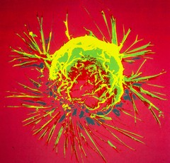
| Description: | A breast cancer cell, photographed by a scanning electron microscope, which produces a 3-dimensional images. The overall shape of the cell's surface at a very high magnification. Cancer cells are best identified by internal details, but research with a scanning electron microscope can show how cells respond in changing environments and can show mapping distribution of binding sites of hormones and other biological molecules. |
| Topics/Categories: |
Cancer Types -- Breast Cancer Cells or Tissue -- Abnormal Cells or Tissue |
| Type: | Color, Photo (JPEG format) |
| Source: | National Cancer Institute |
| Creator: | Bruce Wetzel and Harry Schaefer (Photographers) |
| AV Number: | AV-8000-0302-A |
| Date Created: | 1980 |
| Date Added: | January 1, 2001 |
| Reuse Restrictions: | None - This image is in the public domain and can be freely reused. Please credit the source and, where possible, the creator listed above. |